Ultrasound has improved society in many ways with its medical purposes. Coming from the same technology used in war, sonar has proved to help society immensely in hospitals. It helps pregnant women find problems with their children or give them the choice of knowing the sex of their child, as well as many other problems. Its best feature is the lack of harm it has on the human body and because of this, there are no excuses for it not being used.
Pros
 Governor Scott K. Walker Governor Scott K. Walker Ultrasound in our society consisting of North America (Canada, United states) is praised for the most part. It has no issues that are evident at this moment, and it helps people across these countries immensely. But overseas in other countries, there are some major issues with this technology. In China, ultrasound is heavily watched over and regulated because it is frowned upon to look at the sex of your child before it is born as there is a male dominated preference amongst kids. Parents willingly abort their children if it is a female fetus and their growing population restricts one child per family. This same problem exists in India and the reasoning for the preference of boys over girls is because women's rights are still not seen as equal to those of men in these countries. This can be seen even in North America, but not to the extent of foreign countries. Women earn less and are treated terribly and because of this, they are aborted at birth and ultrasound aids in this. They are calling this issue female feticide. Ultrasound is undoubtedly a great tool in the medical world, but now it is being forced onto women seeking abortions.
In 2013, Governor Scott K. Walker of Wisconsin signed an abortion bill that requires an ultrasound before an abortion. The goal is to form a bond with the mother and unborn fetus in attempts to reconsider the abortion and save the child. Even during the ultrasound, the technician has to tell the patient about the working organs and external features. Depending on the procedure, your doctor could tell you to fast for a number of hours because food in the stomach may interfere with sound waves, and you must notify your doctor of all medications that you are taking at the moment.
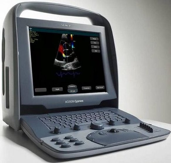
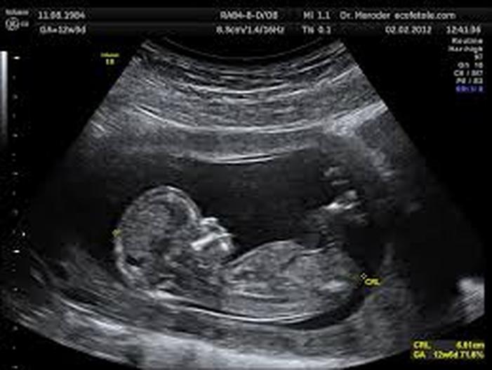
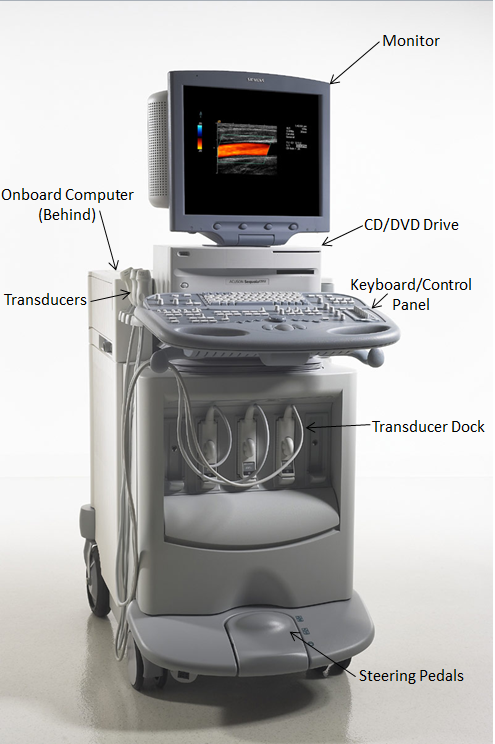
1. The patient will change into a hospital gown so the ultrasound technician has an easier job because clothes restrict access as they are designed to cover the body, the gown might have a cut out for which ever part of the body is being looked at for the day 2. After the patient has changed and is prepared for the ultrasound by following their doctors orders, the sonographer will inform the patient to lay down on an examining table to begin the procedure 3. A jelly like lubricant will be applied to the area where the transducer will be placed because the sonograher does not want friction and it will help transmit the sound waves through the body 4. High frequency sound waves will be produced through the transducer into the area of interest and as the waves travel through your body they will pass through softer tissues as they are designed to, but when they hit a dense object, they will create an echo that will come back to the transducer and be processed and create the image 5. Once the technician is finished, they will clean up the area and the patient will have the freedom to do whatever they please with no restrictions Science An ultrasound can send about one to five megahertz through the body and this technology is closely related to radar and sonar. A frequency or wave of sound is sent out and passes through soft tissues in the body in this case and keeps going until it hits a dense object that bounces the sound wave back to the object that initially sent out the wave; the transducer. The machine calculates each time an echo is sent out and retrieved and this provides the distance between the transducer and the specified tissue/organ and then a 2-D image is created that shows what was in between the transducer and the boundaries which would be a denser object in the body. An ultrasound is a machine used by a sonographer (ultrasound specialist) to look into the human body, specifically areas of soft tissue. It is highly used during pregnancies to view the developing fetus. Ultrasound is also used to see the liver, gallbladder, lymph nodes, ovaries, testes, and many other parts of the body. The parts that make up the ultrasound are the probe, a central processing unit, transducer pulse controls, a monitor, keyboard, a storage device of some sort, and finally a printer. Ian Donald introduced the ultrasound in 1956 but many attempts and references were made before by several people. For example, a man named Galton created and produced a machine that was able to reach 40.000 GHz in 1880. Donald first used ultrasound to measure the diameter of a fetal head and was successful in doing so. He went on to develop this machine that is so widely used today with another man named Brown.
|